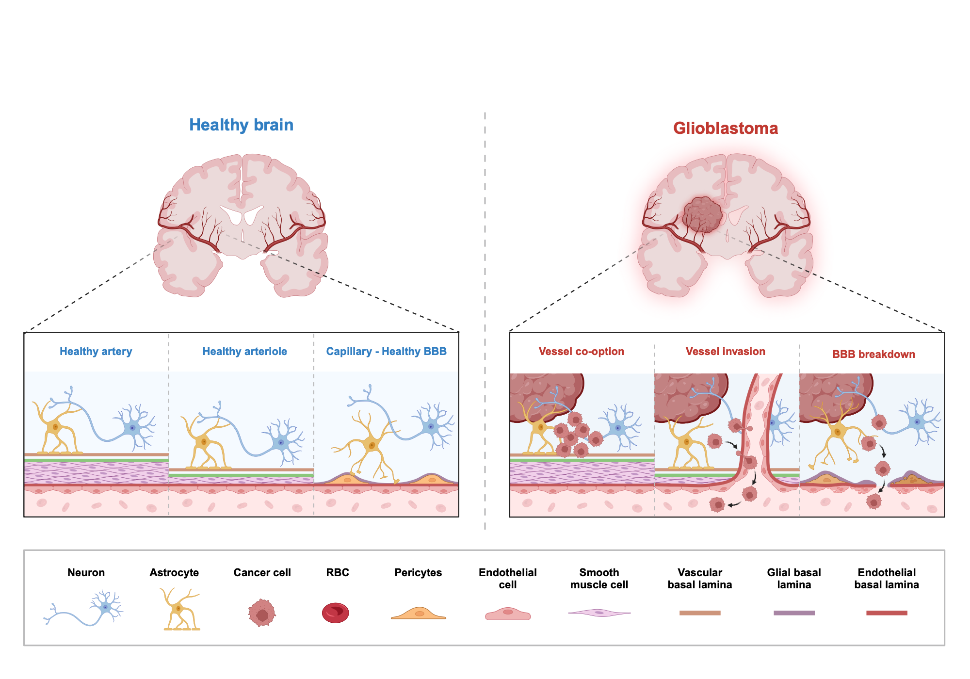

Neuropathological Illustration Comparing Vascular Structures in a Healthy Brain Versus Glioblastoma Invaded Brain Tissue The schematic representation illustrates the differences between normal brain vasculature and the pathological vascular microenvironment in glioblastoma. In a normal brain, the blood-brain barrier (BBB) provides a protective layer that keeps unwanted components out of the brain tissue. This barrier involves the cooperation of several key cells, including neurons, astrocytes, pericytes and endothelial cells. The left column depicts a normal brain with preserved blood-brain barrier (BBB) structures where pericytes, astrocytes and endothelial cells maintain healthy arteries, arterioles and capillaries supported by neurons. A glioblastoma-invaded brain is depicted on the right panel, with vessel co-option (black arrow), vessel invasion (white arrows) and breakdown of the BBB as hallmarks of cancer progression. Some of these alterations in pathology enable infiltration into surrounding brain tissue and destruction of the blood–brain barrier (BBB); both allow for expansion and dissemination of glioblastoma. By contrast, in glioblastoma, cancer cells derange this organization via vessel co-option (tumor cells use pre-existing blood vessels), followed by infiltration of tumor cells into the vascular wall, and ultimately BBB disruption. These adaptations allow cancer cells to metastasize, modify blood circulation, and provide more nutrients and oxygen to the tumour. The BBB dysfunction hampers effective treatment delivery by hindering therapeutic agents from reaching to tumor site, which makes glioblastoma aggressive phenotype and resistant behavior towards conventional therapies.