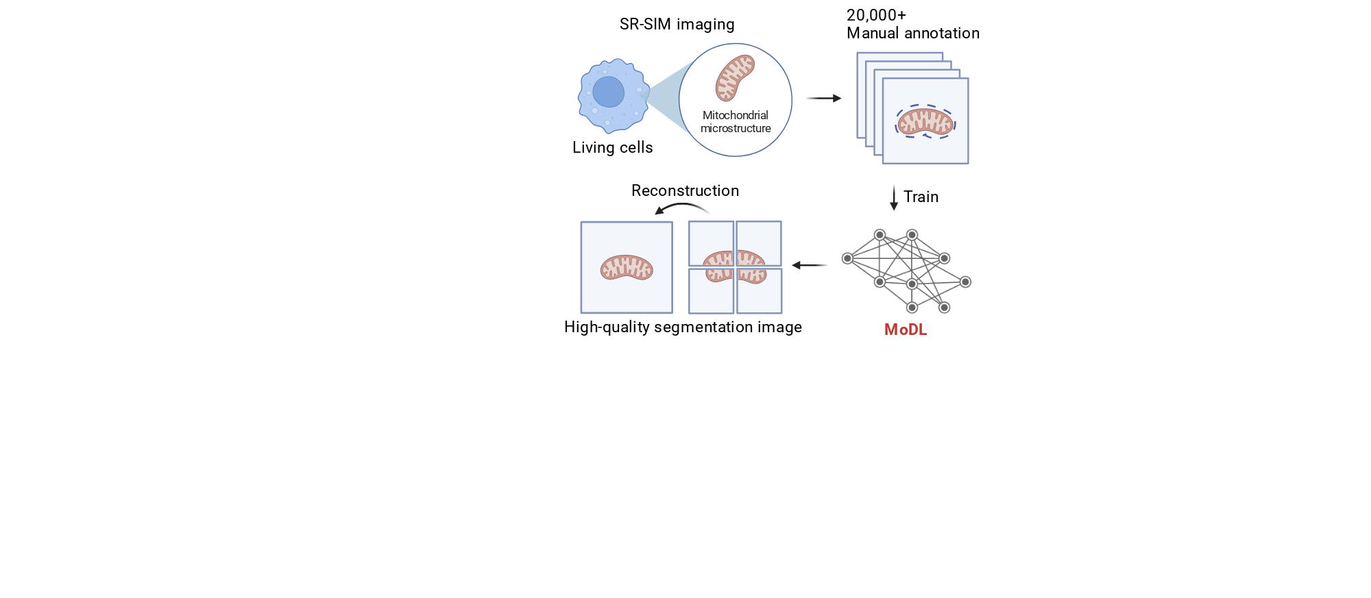
Fig. 2a. Schematic diagram showing super resolution-structured illumination microscopy (SR-SIM) imaging and the key steps of MoDL in mitochondrial segmentation.
Created in BioRender. Li, L. (2024) https://app.biorender.com/citation/673c040df1cf60168c7aaefe
Copy bibliographic reference (APA Style)
Li, L. (2024). Fig. 2a. Schematic diagram showing super resolution-structured illumination microscopy (SR-SIM) imaging and the key steps of MoDL in mitochondrial segmentation.. Created in BioRender. https://app.biorender.com/citation/673c040df1cf60168c7aaefe