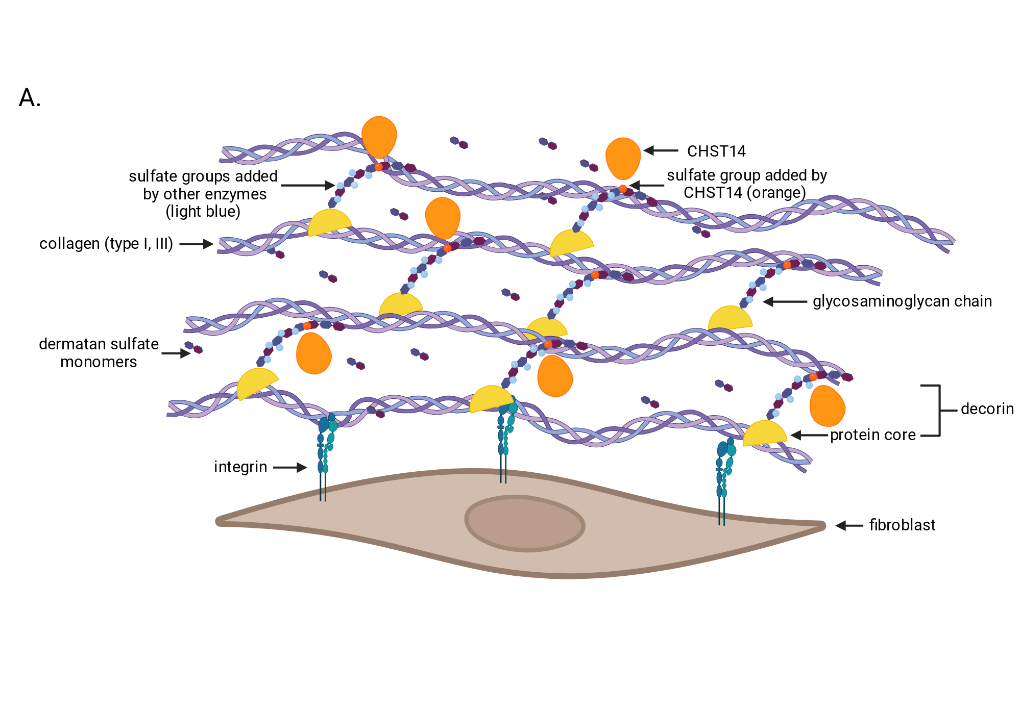
Figure 1H. Model of normal extracellular matrix between cells in the Achilles tendon area shows an organized matrix that promotes normal development of the Achilles tendon.
Created in BioRender. Gumienny, T. (2025) https://app.biorender.com/citation/675a256ce4669bc39b121a29
Copy bibliographic reference (APA Style)
Gumienny, T. (2025). Figure 1H. Model of normal extracellular matrix between cells in the Achilles tendon area shows an organized matrix that promotes normal development of the Achilles tendon.. Created in BioRender. https://app.biorender.com/citation/675a256ce4669bc39b121a29