Figure 6. Experimental design

eye open icon
View-only
chevron left icon
chevron right icon
chevron left icon
chevron right icon
No items found.

This figure is created in BioRender. Create your own professional science figure now!

Explore BioRender’s library of over 50,000 pre-made icons and templates from more than 30 fields of life sciences.
Try it now
Published figure
Link to publication coming soon
View in Mdpi
chevron right icon

Description

No description has been provided for this figure.

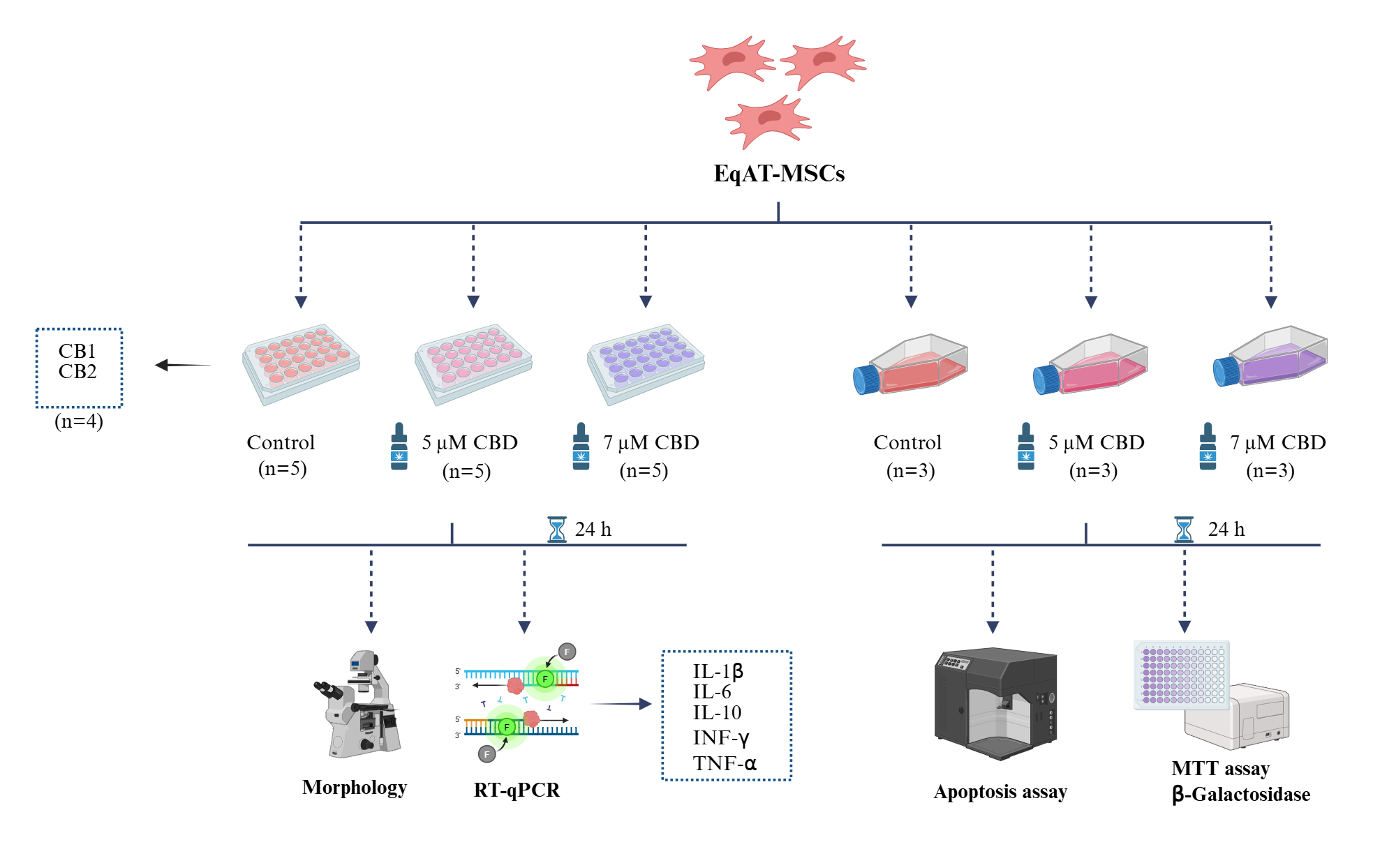
Equine adipose tissue-derived mesenchymal stem cells (EqAT-MSCs) (P3) were primed with 5 µM of cannabidiol (CBD)-rich cannabis extract + complete culture medium and the other group was primed with 7 µM of CBD-rich cannabis extract + complete culture medium. EqAT-MSCs cultured in a complete culture medium were used as the control. After 24 h of priming, the morphological evaluation and gene expression analysis of cytokines such as interleukin 1 beta (IL-1β), interleukin 6 (IL-6), interleukin 10 (IL-10), interferon-gamma (IFN-γ), and tumor necrosis factor-alpha (TNF-α) were performed. Cell metabolic activity was assessed using the MTT assay, and cellular senescence was evaluated through β-galactosidase activity. The apoptosis assay was carried out using annexin and 7-AAD. In addition to the evaluation of cannabinoid receptors 1 (CB1) and 2 (CB2) gene expression in naïve EqAT-MSCs (P2–P5).

Confirmation of the Author’s Publication and Licensing Rights

No confirmation has been provided for this figure.

How to cite this figure

text snippet icon
Copy in-text citation
Created in BioRender. Amorim, R. (2025) https://app.biorender.com/citation/6745ff5939d174eac4087843
text snippet icon
Copy bibliographic reference (APA Style)
Amorim, R. (2025). Figure 6. Experimental design. Created in BioRender. https://app.biorender.com/citation/6745ff5939d174eac4087843

How to use this figure

Show More
Show Less