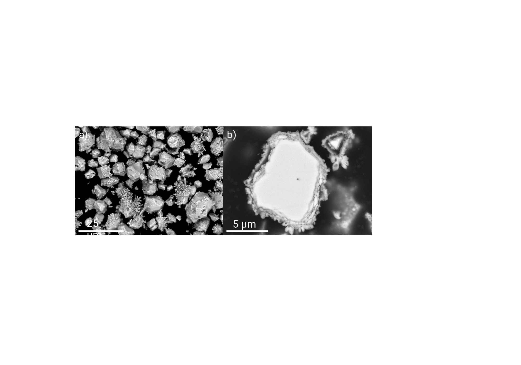
Figure 6: SEM BSE images of the Cu deposit on powdered Nd-Fe-B electrode, i.e., the Nd2Fe14B grains at E= -0.5 V for 30s (a) and on single Nd2FeB14 grains in cross-section at E= -0.5 V for 30s (b).
Created in BioRender. Sodnik, N. (2024) https://app.biorender.com/citation/6708fb9420ac3e30408be1f2
Copy bibliographic reference (APA Style)
Sodnik, N. (2024). Figure 6: SEM BSE images of the Cu deposit on powdered Nd-Fe-B electrode, i.e., the Nd2Fe14B grains at E= -0.5 V for 30s (a) and on single Nd2FeB14 grains in cross-section at E= -0.5 V for 30s (b).. Created in BioRender. https://app.biorender.com/citation/6708fb9420ac3e30408be1f2