Figure 8. Proposed Algorithm for the Evaluation of Cardiac Involvement in EGPA

eye open icon
View-only
chevron left icon
chevron right icon
chevron left icon
chevron right icon
No items found.

This figure is created in BioRender. Create your own professional science figure now!

Explore BioRender’s library of over 50,000 pre-made icons and templates from more than 30 fields of life sciences.
Try it now
Published figure
Link to publication coming soon
View in
chevron right icon

Description

No description has been provided for this figure.

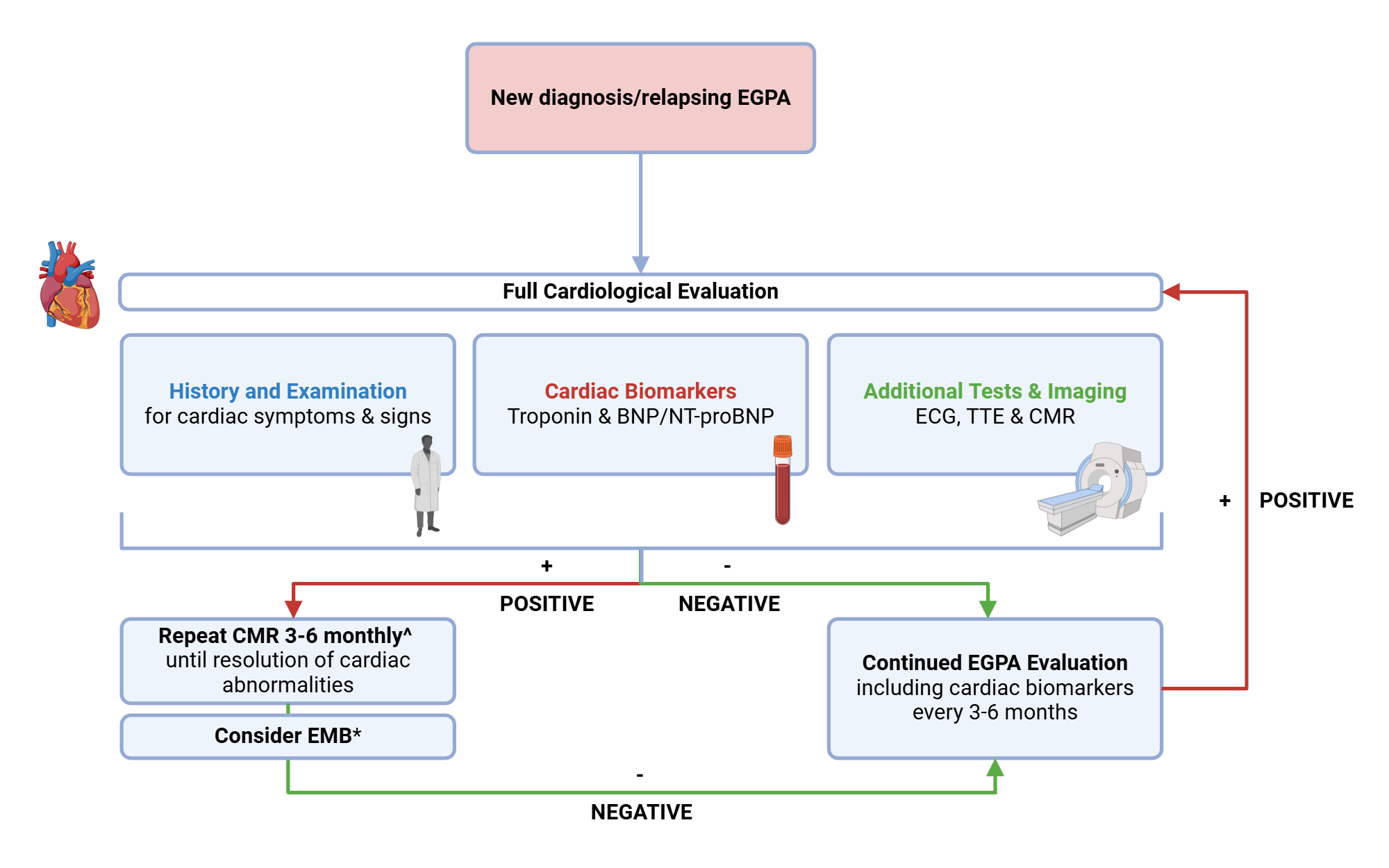
Figure 8. Proposed Algorithm for the Evaluation of Cardiac Involvement in EGPA.^In our experience, 3-6 months is an appropriate time interval to detect improvements in inflammation on CMR. Frequency of further surveillance imaging should be tailored to the individual and requires the input of the multidisciplinary team. Cardiac-gated FDG-PET may be considered as an alternative/adjunctive diagnostic imaging technique in those who have contraindication/intolerance to CMR, or in those in whom uncertainty remains.*Consider EMB for histopathological diagnosis dependent on patient preference and local expertise.BNP, B-type natriuretic peptide; CMR, cardiac magnetic resonance; ECG, electrocardiogram; EGPA, eosinophilic granulomatosis with polyangiitis; EMB, endomyocardial biopsy; NT-proBNP, N-terminal B-type natriuretic peptide; TTE, transthoracic echocardiography.

Confirmation of the Author’s Publication and Licensing Rights

No confirmation has been provided for this figure.

How to cite this figure

text snippet icon
Copy in-text citation
Created in BioRender. Wilson-Morkeh, H. (2025) https://app.biorender.com/citation/67ddca9139af971fc5f23053
text snippet icon
Copy bibliographic reference (APA Style)
Wilson-Morkeh, H. (2025). Figure 8. Proposed Algorithm for the Evaluation of Cardiac Involvement in EGPA. Created in BioRender. https://app.biorender.com/citation/67ddca9139af971fc5f23053

How to use this figure

Show More
Show Less