The anatomy of nociception and pain perception in the cat and dog

eye open icon
View-only
chevron left icon
chevron right icon
chevron left icon
chevron right icon
No items found.

This figure is created in BioRender. Create your own professional science figure now!

Explore BioRender’s library of over 50,000 pre-made icons and templates from more than 30 fields of life sciences.
Try it now
Published figure
Link to publication coming soon
View in Frontiersin
chevron right icon

Description

No description has been provided for this figure.

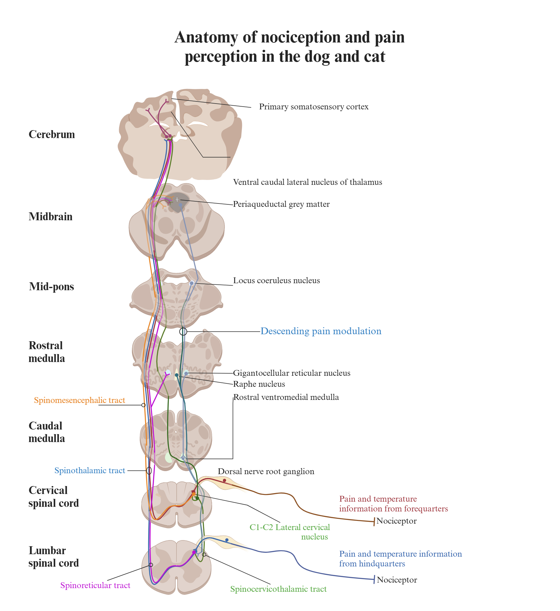
Schematic diagram of the anatomy of nociception and pain perception in the cat and dog. The pathway from registering noxious stimuli to perceiving pain involves multiple nociceptive tracts from various regions of the dorsal horn. Each tract transmits different sensory inputs to specific brain regions, contributing to the perception of pain based on a complex set of input. The spinocervicothalamic and spinothalamic tract are contributing to conscious pain perception, as they relay information to higher brain structures via the thalamus, whereas the spinoreticular and spinomesencephalic tract terminates in the brainstem and the reticular formation and plays a role in the descending modulatory system.

Confirmation of the Author’s Publication and Licensing Rights

No confirmation has been provided for this figure.

How to cite this figure

text snippet icon
Copy in-text citation
Created in BioRender. Pedersen, T. (2025) https://app.biorender.com/citation/67406f3edad780c6b2a5447e
text snippet icon
Copy bibliographic reference (APA Style)
Pedersen, T. (2025). The anatomy of nociception and pain perception in the cat and dog. Created in BioRender. https://app.biorender.com/citation/67406f3edad780c6b2a5447e

How to use this figure

Show More
Show Less